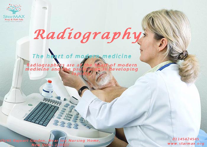
Radiography is the use of X-rays or gamma sun rays to measure non-uniformly made up material; images are registered on a sensitized surface, such as photographic film or a digital detecting. Medical radiography includes an examination of any part of the body for diagnostic purposes, such as detecting fractures or a bowel obstruction. In radiography, the process to produce a picture is quite different. The camera is actually a radiation source and it operates quite totally different to what would be the tradition a photographic camera. The film is not located inside the camera but instead is located on the opposite side of the thing being imaged. The radiation hasn't reflected the film, but instead goes through the object and then strikes the film. The image on the film is dependent after how most of the radiation makes it through the item also to the film. Some materials like bone and metal stop more of the radiation from passing through than do materials like flesh and plastic. The amount of material that the X-rays must traverse also influences how many X-rays reach the film. Differences in the sort of material and the amount of material that the X-rays must enter are in charge of the details in them.
X-Ray is the hospital's first impression of a patient. The same as first impressions with people, the first image used helps set the way going forward. With our start-up mentality, we're centered on reinventing X-Ray to be an intuitive and technologically powerful tool that helps you deliver better confidence. Our mind is set on helping you swiftly and carefully determine the right course of action to condition amazing and valuable care -- all from that first image.
Type of Radiography: -
X-ray (Radiography) Generally there are three types of diagnostic radiographs consumed in this dental offices -- periapical (also known as intraoral or wall-mounted), panoramic, and cephalometric.
Periapical radiographs are probably the most familiar, with images of a few teeth at a time captured on small film cards inserted on the teeth. Periapical ray x machines are normally mounted on the wall inside each treatment room.
Panoramic ("pan") x-rays generate a 5" x 11" (or 12-15 cm x 30 cm) wrap-around radiographic picture of the patient's mouth. This is certainly useful for studying the patient's jaw and the position of the tooth relative to one another. As previously mentioned, there are many additional locations of the patient's structure that can be imaged with a panoramic machine. The pan usually uses up its own small niche in the dental office. Nevertheless, many offices have dedicated x-ray rooms where the machine is located.
Cephalometric ("ceph") x-rays catch a radiographic picture of the complete head, usually in profile. These films are generally employed by orthodontists to diagnose misalignment of the jaw and bite problems. Ceph images are used on a standard beautiful machine outfitted with a cephalometric film-holding arm installed off to one aspect.
A Few Words Regarding Digital Radiography Conventional radiographs are taken on photo taking style film, which must be chemically developed. Technology now offers dentists another option -- digital radiography. Digital radiographs are captured electronically, loaded into, seen and stored on the office's main computer system. Digital radiographs can be increased in many ways; enlarged or reduced, colorized, lightened or darkened. Correct measurements can be considered right off the display. Radiographs can be added to computerized patient documents, printed on paper for the person to take home, incorporated into letters or memos, and electronically sent to insurance providers or affiliate dentists. Digital radiography is not only versatile; it also eliminates the costs and space required for darkrooms, film, and refinement chemicals. Radiation levels are substantially reduced (up to 90%), making the treatment safer for the individual and staff. In addition, time, money, and paperwork are saved in storing and transmitting the images in electronic format. With digital radiography, is actually possible for a basic practitioner to e-mail a radiograph to an experienced professional for consultation while his / her patient is still in the chair. Two main types of digital imaging at present exist: indirect and immediate. The doctor can use his or her existing x-ray equipment to take digital radiographs using either method.

